
This is a joint research project between IMAG and CREATIS.
Overview
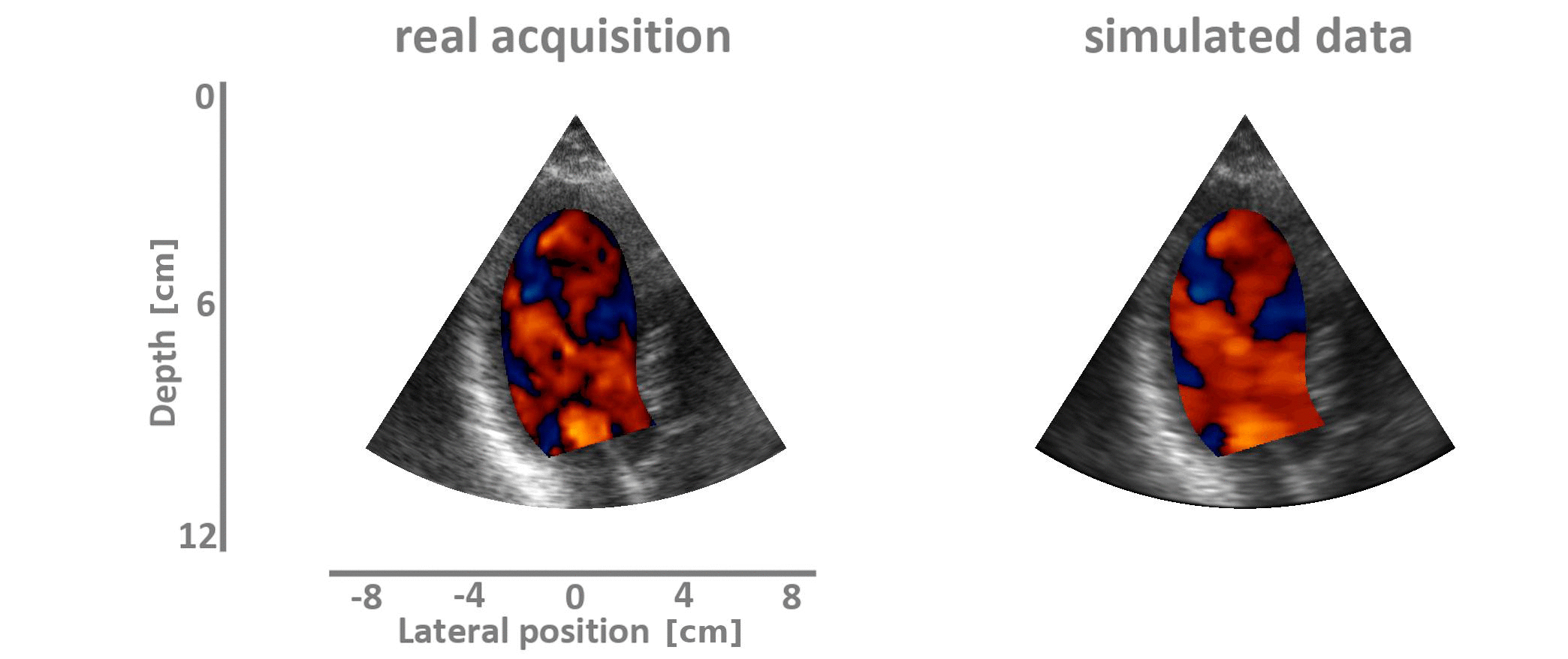
Color Doppler imaging is a modality of choice for simultaneous visualization of myocardium and intracavity flow over a wide scan area. However, this visualization modality is subject to several sources of error, the main ones being aliasing and clutter. Mitigation of these artifacts is a major concern for better analysis of intracardiac flow.
Objectives
we proposed in this study a numerical framework for generating clinical-like color Doppler imaging. Synthetic blood vector fields were obtained from a computational fluid dynamics model. Realistic texture and clutter artifact were simulated from real clinical ultrasound cineloops. We have simulated several scenarios highlighting the effects of i) flow acceleration, ii) wall clutter, iii) transmit wavefronts, on Doppler velocities. As a comparison, an ”ideal” color Doppler, without these harmful effects, was also simulated. This synthetic dataset is publicly available and can be used to evaluate the quality of Doppler estimation techniques.
Methodology
The main steps of our study are as follows :
- the development of the first simulation pipeline for the generation of realistic duplex ultrasound sequences, i.e. the simulation of both B-mode and Doppler images;
- the integration into our simulation of a patient-specific computational fluid dynamics (CFD) model to generate ground-truth blood vector fields;
- the introduction of controlled clutter noise thanks to simple modeling of myocardial movement;
- the assessment of the validity of our pipeline through the simulation of different synthetic duplex sequences.
An overview of the simulated and in-vitro databases is available hereunder.
Open-access database
The dataset corresponds to 20 benchmarked ultrasound duplex sequences composed of 16 B-mode and color Doppler images. The following probe settings were used : a 2.5 MHz 64-element cardiac phased array emitted at a PRF of 7000 Hz. The bandwidths at -6dB were respectively 60% and 20% for the B-mode and Doppler sequences, which corresponded to transmit pulses of 2 and 6 wavelengths. Assuming a heart rate of 60 beats per minute, a focused-beam configuration returned sixteen 11-cm deep B-mode frames interleaved with color Doppler images obtained with packet lengths of 8.
Get Started
To browse through the image database, simply connect to the DUPLEX database, explore and download the images of interest or the entire collection. This database is public, so no login is required.
R&D Team
| Yunyun SUN | PhD student, CREATIS, France
|
| Florian Vixège | PhD student, CREATIS, France
|
| Khuram FARAZ | Postdoctoral fellow, CREATIS, France
|
| Simon MENDEZ | Researcher, IMAG, France
|
| Franck Nicoud | Professor, IMAG, France
|
| Damien GARCIA | INSERM researcher, CREATIS, France
|
| Olivier BERNARD | Associate Professor, CREATIS, France
|